
In our research, we have extensively employed biocompatible semiconductor nanostructures, such as silicon and silicon carbide, to precisely target single cells or specific subcellular components (Table 1). This approach has been instrumental in addressing the limitations of traditional metal electrode-based devices, which are often bulky, disruptive to cell membranes, and reliant on genetic modifications. By integrating these nanostructures at biointerfaces, we successfully identified and quantified their diverse physicochemical outputs, including photo-thermal, photo-faradic, and photo-capacitive effects, establishing a foundation for their use in bioelectronic modulation. These findings demonstrate the remarkable potential of semiconductor-based biointerfaces to modulate electrical activity in various cell types, including neurons, cardiomyocytes, and bacterial cells (Table 1).
Beyond photostimulation, we also explore traditional electrical stimulation to uncover new biological phenomena, demonstrating how semiconductor biointerfaces enable precise, non-invasive modulation across diverse applications. A key example of our work is the discovery of “selective excitability” in Staphylococcus epidermidis, where we used mild electrical stimulation under acidic conditions to suppress bacterial virulence and biofilm formation.
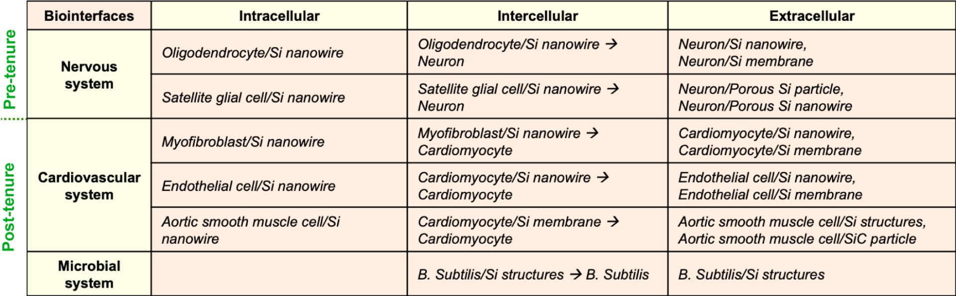
Table 1: Summary of the non-genetic optically-triggered biological modulation
A) Neuromodulation
Neural stimulation methods remain a cornerstone technique in neuroscience. Besides the traditional electrode-based methods and optogenetics, semiconductor-based biomaterial interfaces have enabled wireless, non-genetic, multiscale, high-resolution, random-access photomodulation of neural activities. We have an active research program that aims to study the mechanisms and to validate the efficacy of recently developed nanostructured semiconductor-based neuromodulation tools.
-
Photothermal neuromodulation
My lab set out to demonstrate that silicon’s photothermally induced electric effect could be applied to living cells. We synthesized a deformable and porous type of silicon with molecular-level feature sizes. We introduced the silicon particles over dorsal root ganglia (DRG) neuron cultures and illuminated the cell-membrane-supported particles, eliciting action potentials (signals) in individual neurons (Fig. 1A-B). We also successfully delivered a train of light pulses and repeatedly excited neurons with a one-pulse-one-spike fidelity. This confirmed that the photothermally induced electric effect could indeed be applied to living cells. Silicon’s photothermal effect at the neuron-silicon interface does not require direct physical contact as the heating can be effective for a distance up to one hundred micrometres. This makes it ideal for use in situations such as peripheral nerve stimulations where extracellular matrix or other cellular barriers would usually impede tight biointerfaces.
-
Photoelectrochemical neuromodulation
For greater efficiency in neuromodulation, a tight interface between the silicon device and the neuron is required. When direct access to the cells is available, the preferred neuromodulation approach would be to use electrons and holes (i.e., the charge carriers) that are generated by light, the way a photoelectrochemical device works.
To investigate the biological applicability of silicon’s photoelectrochemical effect, my lab used coaxial p-type/intrinsic/n-type silicon (PINS) nanowires to wirelessly and photoelectrochemically modulate primary rat DRG neuron excitability (Fig. 1C-D). Our results showed that atomic gold on the nanowires enhances the photoelectrochemical process through which the action potentials in rat DRG neurons were elicited. Essentially, atomic gold reduces the kinetic barrier necessary for the photoelectrochemical current generation, thereby playing the role that a catalyst would play in traditional photoelectrochemical devices.
-
Formulation of a rational design principle for semiconductor-based modulation tools
Efficient biological modulation requires accurate designs for tight cell-device interfaces. We identified a biology-guided two-step design principle for establishing tight intra-, inter-, and extra-cellular silicon-based interfaces in which silicon and the biological targets have matched mechanical properties and efficient signal transduction. To gain a biophysical understanding of the different biological modulations that silicon could induce, my lab developed a set of matrices to quantify and differentiate the capacitive, faradaic, and thermal outputs from different silicon materials in saline. We confirmed that we could use light to (non-genetically) modulate intracellular calcium dynamics, cytoskeleton-based transport and structures, and cellular excitability, highlighting the diverse utility of these new interfaces. In particular, we showed that flexible and freestanding silicon mesh can modulate brain activities and simple animal behaviors such as induced limb motion from anaesthetized mice (Fig. 1E-F).
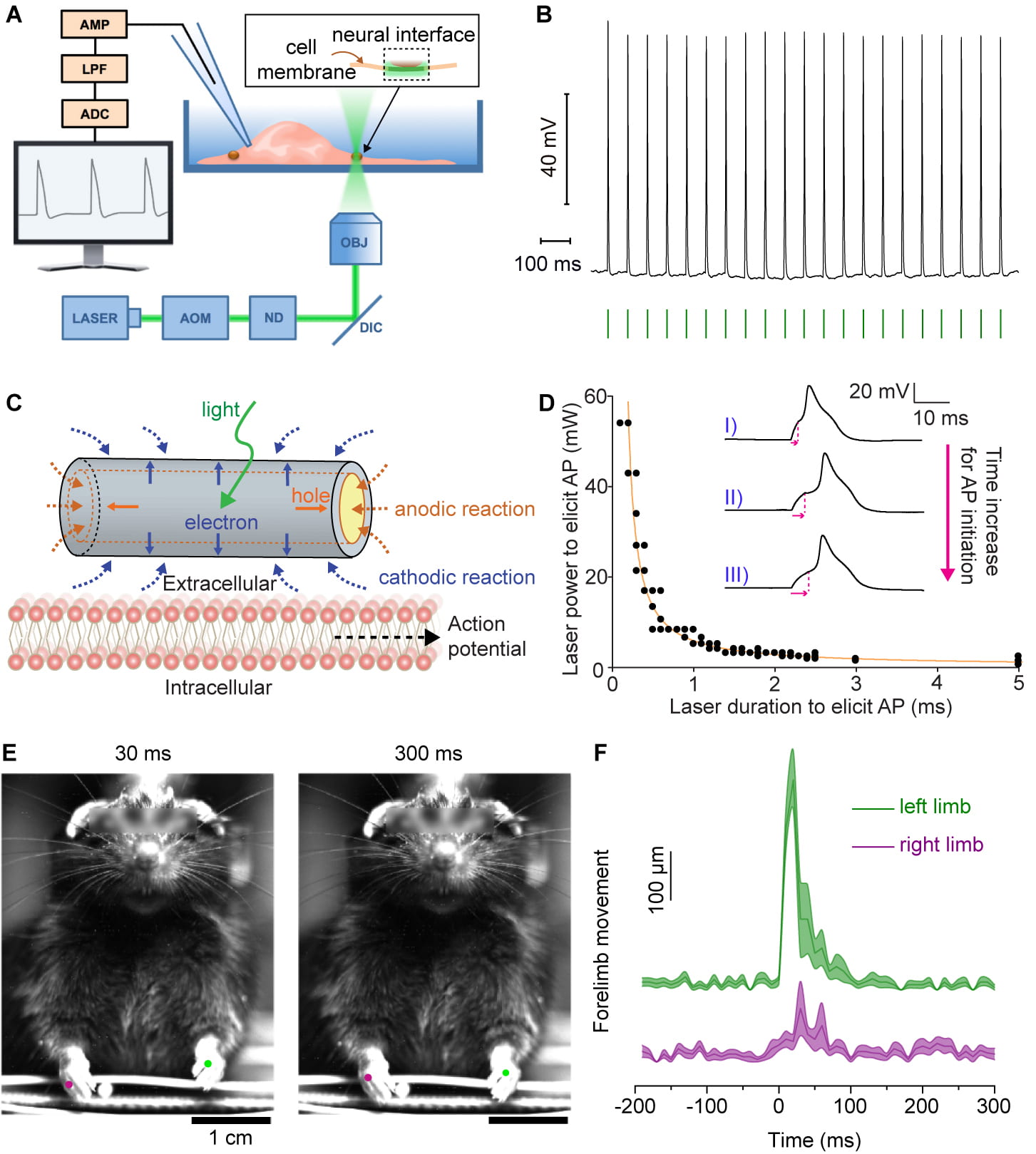
Figure 1. Neuromodulation. (A-B) Nanoporous silicon-based neuromodulation through an optocapacitance mechanism. (C-D) Coaxial silicon nanowire for photoelectrochemical neuromodulation. (E-F) Multilayered silicon membranes for neuromodulation at the animal level.
B) Cardiac modulation
The electrical conduction system of the heart allows for the coordinated contraction of cardiomyocytes to produce heartbeats. Abnormalities in this system can lead to delayed mechanical activation of specific regions of the heart or pathologically slow heart rates (bradyarrhythmias). Thus, therapies that can either resynchronize the heart or increase the overall beating frequency of the heart are necessary for the treatment of these disorders.
-
Extracellular cardiac modulation
The Tian lab recently developed a photoelectrochemical method to optically modulate cardiac beating to a specified target frequency in primary cultured cardiomyocytes and adult rat hearts ex vivo (Fig. 2). To achieve this modulation, we used a low irradiance moving laser stimulus and a biocompatible polymer-silicon nanowire composite material (Fig. 2A). We integrated the PINS nanowires, initially developed for neuromodulation, with polymeric mesh, creating a composite sheet for conformal cardiac interfaces both in vitro (Fig. 2B) and ex vivo (Fig. 2C-D). Upon light stimulation, the PINS nanowires produce photoelectrochemical output, which depolarizes cardiomyocyte plasma membrane and eventually leads to the training or pacing effect. This work has implications for future bioelectrical studies of the cardiac conduction system as well as therapeutics for cardiac conduction disorders in the clinic.
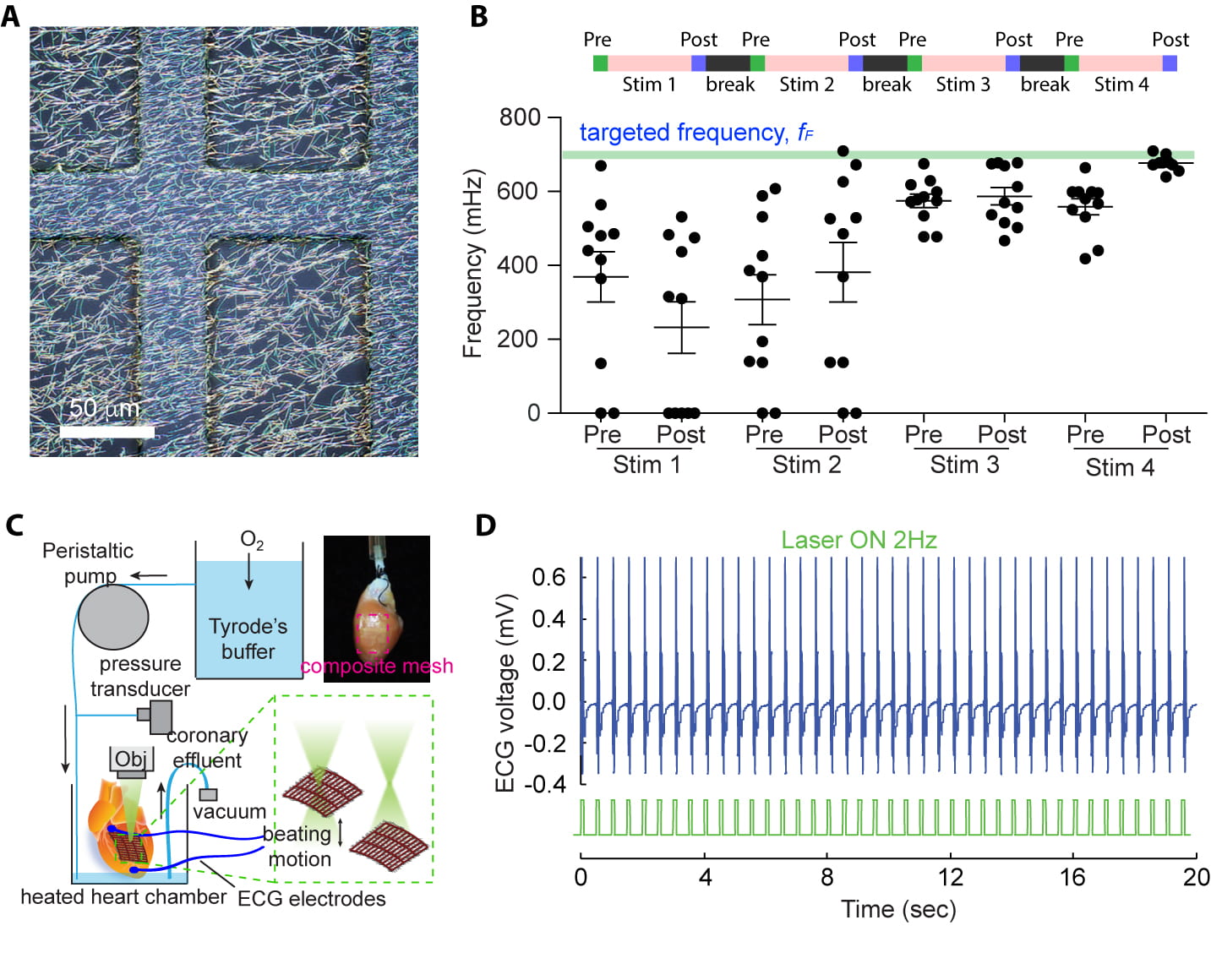
Figure 2. Extracellular cardiac optical modulation with polymer/Si nanowire composite mesh. (A) Optical microscopy image of the composite. (B) Optical training in vitro, showing gradual increase in the cardiac beating frequency. (C) Schematic diagram for optical training and pacing in vivo. (D) ECG recording, showing the optical pacing with the composite mesh.
Recently, we presented a significant innovation in optoelectronic devices for biological modulation (Fig. 3). We introduced a porosity-based heterojunction, created in p-type silicon. This novel approach marks a departure from conventional methods that rely on materials with varying doping or composition, which are often expensive and complex to fabricate. Our methodology involves a blend of stain etching and oxygen plasma treatment to craft optoelectronic devices from bulk silicon. This approach is markedly simpler and more direct than traditional fabrication methods, such as chemical vapor deposition. It enables us to produce devices capable of modulating biological tissues using low optical power densities and near-infrared light. Our findings show that these stain-etched heterojunctions can generate robust photocurrents upon light illumination, significantly boosted by oxygen plasma treatment. This enhancement is attributed to increased hydrophilicity and reduced surface recombinations. Electrochemical tests demonstrated improved capacitance and reduced impedance of the heterojunctions, signaling efficient charge injection and ion transfer capabilities, distinct from traditional gold-decorated p–i–n junctions. We also found that the photoelectrochemical currents depend on the power and wavelength of the illumination light, responding to visible and near-infrared light but not to ultraviolet light. Finally, we demonstrated the application of these devices in modulating isolated cardiac tissue and in vivo sciatic nerve biomodulation. Our devices can interface with biological tissues without adhesives or physical modifications, responding to light-induced stimuli with varying intensity-dependent effects. The simplicity and efficiency of this fabrication approach, coupled with the use of biocompatible p-type silicon, make our porosity-based heterojunction a promising candidate for future bioelectronic therapies, with significant potential for diverse therapeutic applications.

Figure 3. We introduce porosity-based heterojunctions in p-type silicon for bioelectronics, offering a simpler, cost-effective alternative to traditional methods and enabling efficient, leadless optoelectronic stimulation of biological tissues. Our innovative approach, combining stain etching and oxygen plasma treatment, facilitates the development of flexible bioelectronic devices with significant potential in cardiac and nerve modulation, and broad applications in bioelectronic therapies.
-
Intracellular cardiac modulation
The current material tool kit for cardiac modulation is primarily based on synthetic or nonliving components. When interfacing with live and dynamically changing tissues, seamless integration of the device is limited by the remaining mechanical invasiveness of the materials and non-natural biological signal transduction at the biointerface. The Tian lab recently proposed that a living bioelectrical system with dynamic and developing behaviors can advance bioelectrical interfaces due to the adaptability and motility of the cellular components and the diverse physical properties of the materials components. This approach is fundamentally different from biological pacemakers, which are generated by transferring genes that encode transcription factors to transform working myocardium into a surrogate sinoatrial node.
We achieved the living bioelectrical system by intracellular integration of silicon nanowires with myofibroblasts through phagocytosis (Fig. 4A). We then demonstrated that this living hybrid tool can be used to investigate intercellular electrical coupling in vitro and in vivo. Using the myofibroblast-nanowire hybrid tool to compare myofibroblast-myofibroblast electrical coupling with myofibroblast-cardiomyocyte coupling in vitro, we detected two different calcium flux propagation mechanisms – one for amplified cardiomyocytes propagation and the other for passive myofibroblasts propagation. We found that, unlike bare silicon nanowires, the myofibroblasts-silicon nanowire hybrids can be seamlessly integrated into contractile cardiac tissue (Fig. 4).
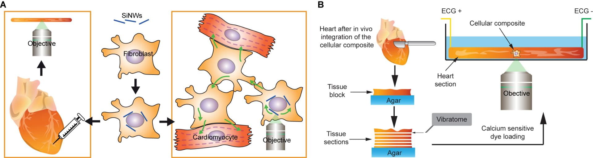
Figure 4. Myofibroblast/Si nanowire composites for intracellular modulation of cardiac activities. (A) The cellular composites are prepared and then used for bioelectrical studies in vitro and in vivo. (B) Procedure for bioelectrical studies ex vivo.
C) Microbial modulation
Bacteria exhibit multicellular-like behaviors through intercellular communication and cooperation. However, their responses to transient physical perturbations, such as thermal or mechanical shocks, remain poorly understood, primarily due to the limitations of traditional tools in capturing rapid, localized microbial responses. Optical modulation techniques, leveraging non-genetic transducers, offer a promising alternative by delivering transient, multiplexed, and spatially precise stimuli to microbial systems (Fig. 5).
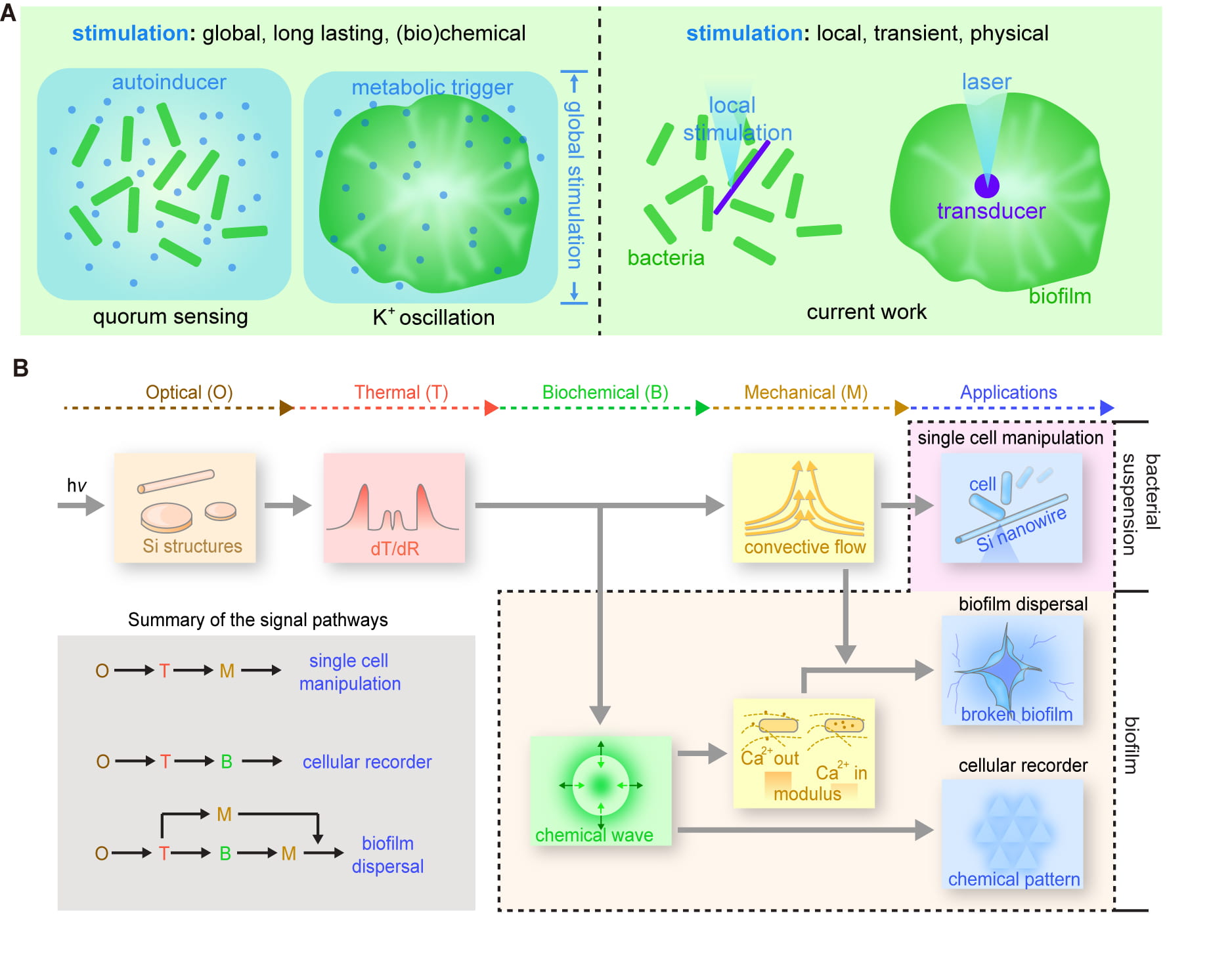
Figure 5. Si-based optical modulation microbial activities. (A) Our approach uses local, transient and physical stimulations. (B) We discovered that thermal gradient can elicit calcium wave in biofilms, which leads to a range of applications such as biofilm dispersal.
Recent breakthroughs have shifted focus toward bacterial electrical excitability, revealing its potential for precise, antibiotic-free bioelectronic control. In particular, our lab uncovered a novel phenomenon termed “selective excitability,” where certain bacteria, such as Staphylococcus epidermidis, exhibit electrical responsiveness only under specific environmental conditions, such as the acidic pH of healthy skin (Fig. 6). At pH 5, these bacteria become electrically excitable, allowing for reversible changes in membrane potential and intracellular pH without inducing cell death. This excitability is driven by a strong transmembrane proton gradient, which is critical for proton influx during electrical stimulation.
Leveraging selective excitability, we developed programmable electrical stimulation protocols that suppress bacterial growth, reduce virulence gene expression, and inhibit biofilm formation. Importantly, these effects were achieved under benign, low-voltage conditions, making this approach safer and more targeted compared to conventional antibiotic treatments. Using this principle, we demonstrated the efficacy of a flexible electroceutical patch for localized control of S. epidermidis colonization on porcine skin models. This work highlights the potential of harnessing selective excitability for sustainable, non-invasive microbial modulation, paving the way for advanced bioelectronic solutions in healthcare and beyond

Figure 6. Mammalian cells like neurons and cardiomyocytes are well-known for their electrical excitability, which is exploited in devices like pacemakers for drug-free therapies. However, the electrical excitability of bacteria remains underexplored despite its potential for addressing antibiotic resistance. In this study, we demonstrated that Staphylococcus epidermidis, a skin-dwelling bacterium, exhibits “selective excitability,” becoming electrically responsive only in the acidic pH of healthy skin. Leveraging this trait, we used mild electrical stimulation to suppress bacterial virulence factors, providing a localized, programmable, and antibiotic-free approach to control opportunistic pathogens.
D) Random-access biological modulation
Our lab has pioneered the development of a random-access, high spatiotemporal resolution, and pixel-less photostimulation platform for translational applications, leveraging monolithic silicon-based photoelectrochemical devices (Fig. 7). These devices address limitations in current clinical and experimental stimulation technologies, such as the spatial constraints of electrode arrays and the challenges associated with optogenetics. By integrating leadless and non-genetic photostimulation capabilities, our platform offers precise control over cellular and tissue-level modulation using light.
The platform employs advanced silicon configurations, including single-crystalline and nanoporous structures, to optimize photocurrent localization and tunability. Photogenerated carriers in these materials enable localized cathodic and anodic processes with millisecond precision, surpassing the spatial resolution of traditional electrode-based systems. This high resolution is achieved through tightly confined carrier diffusion and innovative junction designs, allowing for programmable and site-specific stimulation without the need for pre-designed device patterns.
We have demonstrated the platform’s capabilities across various biological models, from cultured cardiac cells to in vivo pig hearts, showcasing its translational potential. Key achievements include optical pacing of isolated rat hearts using millisecond light pulses and closed-thoracic cardiac modulation in pigs via a custom endoscopic system. These results highlight the system’s ability to perform minimally invasive and multisite photostimulation, effectively addressing complications associated with lead-based devices in cardiac resynchronization therapy.
Beyond cardiac applications, this platform is adaptable for broader translational purposes, including neurostimulation and tissue engineering. By combining high spatial resolution, random-access capabilities, and a lightweight, leadless design, our photostimulation system establishes a versatile framework for advancing bioelectronic therapies in clinical and experimental settings. This work underscores the potential of semiconductor-enabled biointerface.

Figure 7. The design and application of monolithic photoelectrochemical devices for high-resolution bioelectrical modulation. These devices enable localized photostimulation with spatially resolved cathodic and anodic processes, delivering precise, fast, and balanced temporal responses. Advanced imaging reveals innovative semiconductor junction designs optimized for superior photoresponse. Demonstrated across multiscale cardiac models, from neonatal cardiomyocytes to in vivo pig hearts, these devices showcase their adaptability for mechanistic studies and translational therapies, bridging experimental innovation with clinical potential.
Selected Publications
- Y. W. Jiang, J. L. Carvalho-de-Souza, R. C. S. Wong, Z. Q. Luo, D. Isheim, X. B. Zuo, A. W. Nicholls, I. W. Jung, J. P. Yue, D.-J. Liu, Y. C. Wang, V. De Andrade, X. H. Xiao, L. Navrazhnykh, D. E. Weiss, X. Y. Wu, D. N. Seidman, F. Bezanilla, B. Z. Tian, Heterogeneous silicon mesostructures for lipid-supported bioelectric interfaces, Nature Materials, 2016, 15, 1023–1030. Link
- J. F. Zimmerman, R. Parameswaran, G. Murray, Y. C. Wang, M. Burke, B. Z. Tian, Cellular uptake and dynamics of unlabeled free standing silicon nanowires, Science Advances, 2016, 2, e16010139. Link
- R. Parameswaran, J. L. Carvalho-de-Souza, Y. W. Jiang, M. Burke, J. F. Zimmerman, K. Koehler, A. Phillips, J. Yi, E. Adams, F. Bezanilla, B. Z. Tian, Photoelectrochemical modulation of neuronal activity with free-standing coaxial silicon nanowires, Nature Nanotechnology, 2018, 13, 260-266. Link
- Y. W. Jiang, X. J. Li, B. Liu, J. Yi, Y. Fang, F. Y. Shi, X. Gao, E. Sudzilovsky, R. Parameswaran, K. Koehler, V. Nair, J. P. Yue, K. H. Guo, Y. Fang, H.-M. Tsai, G. Freyermuth, R. C. S. Wong, C.-M. Kao, C.-T. Chen, A. W. Nicholls, X. Y. Wu, G. M. G. Shepherd, B. Z. Tian, Rational design of silicon structures for optically-controlled multiscale biointerfaces, Nature Biomedical Engineering, 2018, 2, 508-521. doi:10.1038/s41551-018-0230-1. Link
- R. Parameswaran, B. Z. Tian, Rational design of semiconductor nanowires for functional subcellular interfaces. Accounts of Chemical Research, doi: 10.1021/acs.accounts.7b00555. 2018. Link
- Y. W. Jiang, B. Z. Tian, Inorganic semiconductor-enabled bioelectronic and biophotonic interfaces. Nature Reviews Materials, 2018, 3, 473-490. Link
- P. Parameswaran, K. Koehler, M. Rotenberg, M. Burke, J. Kim, K.-Y. Jeong, B. Hissa, M. Paul, K. Moreno, N. Sarma, T. Hayes, E. Sudzilovsky, H.-G. Park, B. Z. Tian, Optical stimulation of cardiac cells with a polymer-supported silicon nanowire matrix. Proc. Natl. Acad. Sci. USA, 2019, 116, 413-421. Link
- A. Prominski, J. Y. Shi, P. J. Li, J. P. Yue, Y. L. Lin, J. Park, B. Z. Tian, M. Y. Rotenberg. Porosity-based heterojunctions enable leadless optoelectronic modulation of tissues. Nature Materials, 2022, DOI: 10.1038/s41563-022-01249-7. Link
- Y. W. Jiang, R. Parameswaran, X. J. Li, J. L. Carvalho-de-Souza, X. Gao, L. Y. Meng, F. Bezanilla, G. M. G. Shepherd, B. Z. Tian, Non-genetic optical neuromodulation with silicon-based materials. Nature Protocols, 2019, 14, 1339–1376. Link
- B. Z. Tian, C. M. Lieber, Nanowired bioelectric interfaces. Chemical Reviews, 2019, 119, 9136-9152. Link
- H. Acaron Ledesma, X. Li, J. L. Carvalho-de-Souza, W. Wei, F. Bezanilla, B. Z. Tian, An atlas of nano-enabled neural interfaces. Nature Nanotechnology, 2019, 14, 645-657. Link
- B. Z. Tian, Nongenetic neural control with light, Science, 2019, 365, 457, DOI: 10.1126/science.aay4351. Link
- M. Y. Rotenberg, N. Yamamoto, E. N. Schaumann, L. Matino, F. Santoro, B. Z. Tian, Living myofibroblast-silicon composites for probing electrical coupling in cardiac systems. Proc. Natl. Acad. Sci. USA, 2019, 116, 22531-22539. Link
- X. Gao, Y. W. Jiang, Y. L. Lin, K.-H. Kim, Y. Fang, J. Yi, L. Y. Meng, H.-C. Lee, Z. Y. Lu, O. Leddy, R. Zhang, Q. Tu, W. Feng, V. Nair, P. J. Griffin, F. Y. Shi, G. S. Shekhawat, A. R. Dinner, H.-G. Park, B. Z. Tian, Structured silicon for revealing transient and integrated signal transductions in microbial systems. Science Advances, 2020, 6, eaay2760. Link
- S. Kim, E. Eig, J. P. Yue, A. Yang, C. Comerci, M. Laune, C. W. Yang, A. Kamath, J. Y. Shi, P. J. Li, Z. Cheng, C. X. Sun, T. T. Guo, V. Tian, G. M. Suel, B. Z. Tian, Bioelectronic drug-free control of opportunistic pathogens through selective excitability. Device, 2024, 2, 11, 100596. Link
- P. J. Li, J. Zhang, H. Hayashi, J. P. Yue, W. Li, C. W. Yang, C. X. Sun, J. Y. Shi, J. Huberman-Shlaes, N. Hibino, B. Z. Tian, Monolithic silicon for high-spatiotemporal translational photostimulation. Nature, 2024, 626, 990–998. Link
Go to: General Research